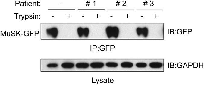
Fig. 4.
IgG4 antibodies from MuSK MG patients do not reduce MuSK cell-surface expression. 3T3 cells, which were transiently transfected with MuSK-GFP, were treated with MuSK MG IgG4 antibodies for 24 h (Fig. 3). Cells were harvested or treated with trypsin before harvesting. MuSK-GFP was immunoprecipitated from lysates and detected in Western blots using antibodies to GFP, and the level of GAPDH in lysates was determined by Western blotting.

