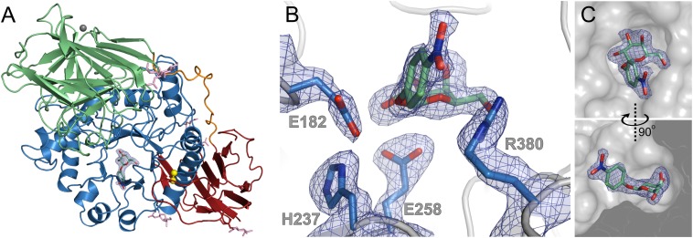
Fig. 2.
Structure of the wild-type GALC enzyme in complex with substrate. (A) Ribbon diagram showing the overall structure of GALC with the substrate 4NβDG bound in the active site. The electron density (2FO-FC contoured at 0.25 e−/Å3, blue) is shown for uncleaved substrate bound in the active site pocket of GALC. Surface glycans (pink sticks) are shown. (B) Detail of the GALC active site with bound, uncleaved substrate showing active site residues (sticks) and electron density (as above). (C) Surface representation of the GALC active site (gray) with the substrate and electron density shown (as above).

