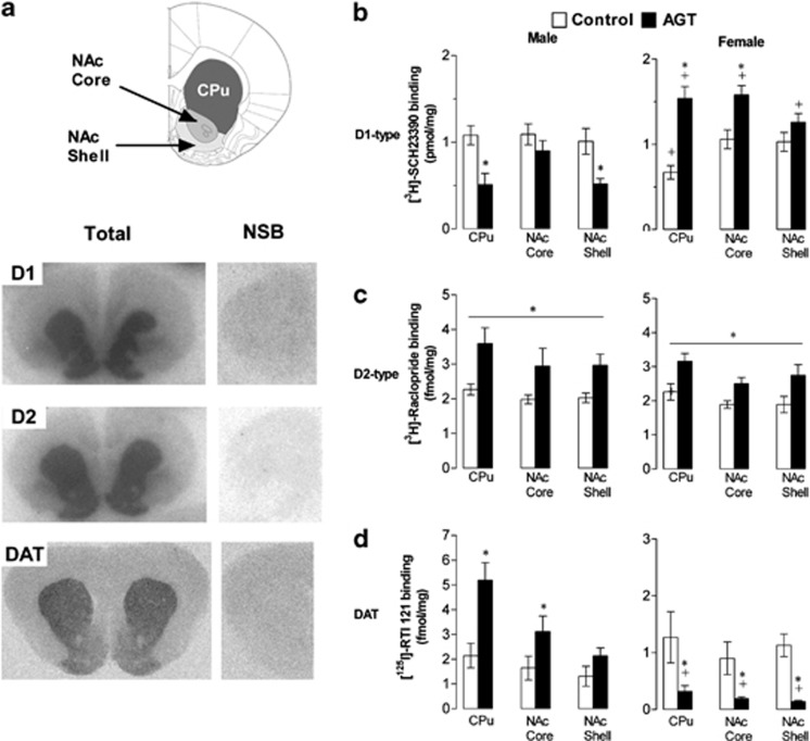
Figure 2.
Sexually dimorphic changes in the expression of dopaminergic (DA) signaling proteins after antenatal glucocorticoid treatment (AGT). (a) Top panel schematically illustrates regions of interest, namely the dorsal striatum (caudate putamen, CPu), nucleus accumbens (NAc) core, and NAc shell based on the rat brain atlas, and a coronal section +2.2 mm relative to bregma (Paxinos and Watson, 1998). Serial sections were collected between +1.7 mm and +2.2 mm relative to bregma for the NAc core and shell, whereas for the CPu serial sections were collected between −0.2 and +1.6 mm relative to bregma. Lower panels are representative autoradiographic images of total (left-hand panels) and non-specific (right-hand panels) ligand binding (NSB) to D1-type receptors (D1), D2-type receptors (D2) and the dopamine reuptake transporter (DA transporter, DAT). (b–d) Quantitative comparison of radioligand binding to D1 (b) D2 (c) and DAT (d) in CPu and NAc core, and shell of male (left-hand panels) and female (right-hand panels) rats exposed to AGT (dexamethasone, 0.5 μg/ml, in drinking water on gestational days 16–19; closed histograms) or untreated controls (open histograms). Note the use of different scales for male and female DAT binding. Data are mean±SEM, n=6, *p<0.05 (post-hoc analysis, (b, d)) vs relative control group, and p<0.001 (c) main effect of AGT and region;+p<0.05 vs corresponding male group, indicating a significant sex difference.

