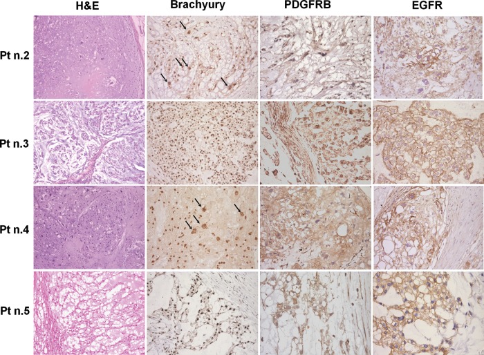
Fig. 2.
Morphology (H&E staining), brachyury and RTK immunohistochemistry findings in the 4 successfully implanted chordomas. All of the cases showed physaliferous cells with brachyury nuclear immunostaining as well as cytoplasmatic PDGFRB and mainly membranous EGFR immunostaining. Patients 2 and 4 showed pleomorphic cells with large polylobated nuclei (indicated by the arrows). Photomicrographs original magnifications; H&E staining and brachyury immunohistochemistry: 50x; PDGFRB and EGFR immunohistochemistry: 100x with the exception of EGFR immunostaining of pt n.5 that is 50X.

