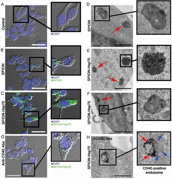
Fig. 4.
Fluorescent and transmission electron microscopy images of C6 glioma cells in vitro. (A) Control C6 cells and (B) cells incubated in slide chambers for 24 h with SPIONs (50 µg/mL) or (C) cells incubated with Hsp70-SPION сonjugates (50 µg/mL) were fixed with 4% PFA, stained with 4′,6′-diamidino-2-phenylindole (DAPI). Images of cell nuclei (blue) and nanoparticles (green) were acquired sequentially. Nanoparticles appear as green dots or larger coarse aggregates inside the cytoplasm. Scale bar, 25 µm. (D) Electron microscopy images of C6 cells incubated with SPIONs (50 µg/mL) for 24 h. Nanoparticles within the cytoplasm of C6 cells as electron-positive inclusions in the membranous structures are shown by red arrow. Scale bar, 1 µm. (E) Hsp70-SPION conjugates presented as electron-positive inclusions in nonmembranous structures and (F) endosomes within the cytoplasm. Scale bar, 1 µm. (G) Fluorescent image of C6 glioma cells incubated with Hsp70-SPION conjugates (50 μg/mL) for 24 h following treatment with blocking anti-CD40 antibodies. Scale bar, 25 μm. (H) Immunocytochemistry of C6 cell stained with anti-CD40 antibodies. The presence of Hsp70-SPION conjugates (red arrows) inside the CD40+ (blue arrows) membrane structures could be shown. Scale bar, 500 nm.

