Fig. 5.
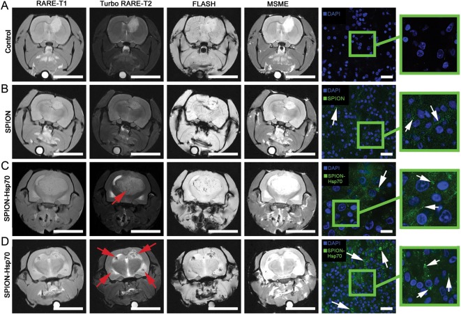
MRIs of (A) C6 glioma for control, (B) SPIONs i.v. injected, (C) Hsp70-SPION conjugates i.v. infused, and (D) the infusion of Hsp70-SPIONs following preliminary injection of Hsp70. Images were taken following 24 h after nanoparticle infusion at different acquisition regimes: RARE-T1, Turbo-RARE-T2, fast low angle shot MRI (FLASH), and multiscan-multiecho (MSME). Scale bar, 1 cm. Red solid arrows point to the zones of “darkening” inside the glioma that correspond to the nanoparticle accumulation. Following assessment on MR scanner, the brains were extracted, dissected, stained by 4′,6′-diamidino-2-phenylindole (DAPI; blue), and analyzed for nanoparticle presence with the help of confocal microscopy. Nanoparticles appear as green dots or larger aggregates (shown by white solid arrows) within the cytoplasm and in the extracellular space. Scale bar, 25 µm.

