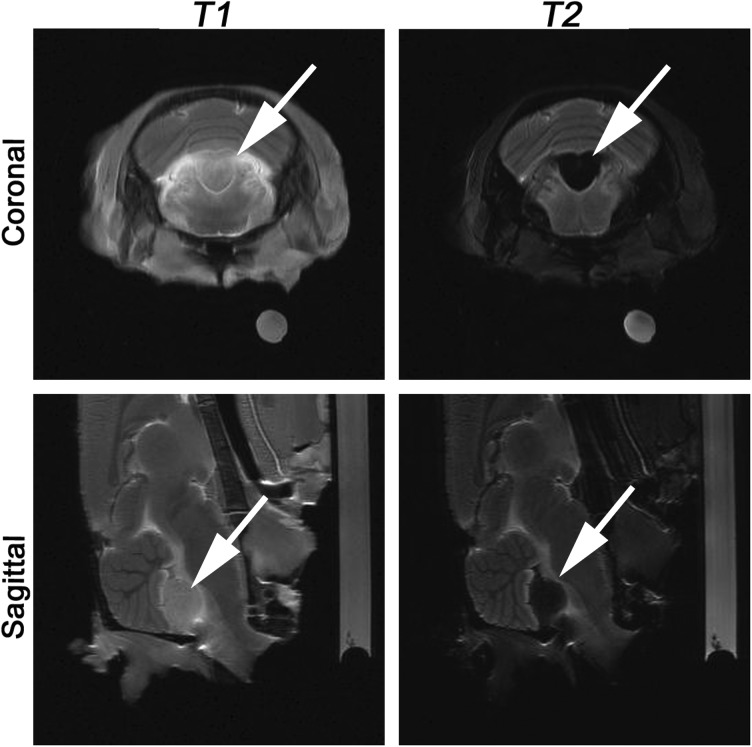
Fig. 6.
MRIs of animal #2. Following i.v. infusion of Hsp70 (5 mg/kg), the Hsp70-SPION conjugates (150 µg/kg) were i.v. injected. Before mounting the animal in the head coil i.v., we injected contrast agent (Gadovist, 1.0 mmol/mL, 1 mL). The T1- and T2-weighted coronal and sagittal brain sections are presented. Red solid arrows point to the location of the C6 tumor. The accumulation of nanoparticles appears as a “dark” core at the T2-weighted regimen. On the Gd-enhanced T1-weighted images, only the slight increase in contrast in the tumor zone is observed.

