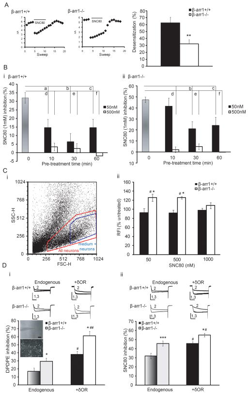
Fig. 2. β-arr1 regulates δOR desensitization and internalization but this does not explain the enhanced δOR-VDCC coupling in β-arr1−/− neurons.
A. Rapid desensitization: Once complete inhibition of VDCCs was obtained by SNC80, further perfusion with SNC80 desensitized this inhibition. This is shown by the decline in the peak current amplitude over 120s in the left panels. Although both β-arr1+/+ and −/− neurons desensitized, β-arr1−/− neurons showed less desensitization, **p<0.01, n=5–10. B. Acute desensitization: Whereas pre-incubation of DRGs with 50 and 500nM SNC80 for 10min desensitized δOR-VDCC inhibition at all time points in +/+ neurons, −/− neurons did not desensitize after 10min of 50nM SNC80 but desensitized thereafter and showed equivalent desensitization at all timepoints when pre-incubated in 500 nM SNC80. β-arr1+/+; a, b, c: p<0.05 vs untreated, d, e: p<0.0001 vs untreated, f: p<0.001 vs untreated, β-arr1−/−; b: p<0.001 vs untreated, c: p<0.001 vs untreated, d, e, f: p < 0.0001 vs untreated, n=6–7 per data point. Columns in shades of grey, Bi; +/+ or Bii; −/−, indicate data shown in Fig 1. C. Internalization assessed by flow cytometry in β-arr1−/− and +/+ DRG neurons using an N-terminal antibody and Allophycocynanin (APC)-conjugated secondary antibody. i The δOR-labeled non-fixed samples were initially sorted by size (FSC-H) and granularity (SSC-H) to select the medium-large DRG neurons (>25 μM in diameter Walwyn et al., 2004). APC-labeled δORs were then selected after removing non-specific background labeling and all samples of each experiment analyzed by these parameters. ii. Although basal cell surface δOR levels were unaffected by the β-arr1−/− deletion, 50 and 500, but not 1000, nM SNC80 increased cell surface δOR levels in βarr1−/− DRGs. #p<0.05 vs untreated β-arr1−/−, * p<0.05 vs β-arr1+/+, same time point, n=4–8 per data point. D. A slower rate of internalization could enhance δOR-VDCC inhibition by increasing the number of δORs on the plasma membrane. This was assessed by over-expressing Cerulean (CFP)-labeled δORs (Walwyn et al., 2009). This increased δOR-VDCC inhibition equally in both β-arr1+/+ and−/− neurons tested with either DPDPE or SNC80 (1μM ea). *p<0.05, **p<0.01 and ***p<0.001 vs β-arr1+/+, #p<0.05, ## p<0.01 vs endogenous δORs, n=6–18 per data point. All exemplar currents depict the current before, during and after agonist application (1, 2 and 3) and the scale bars show time, 20ms, and current, 5nA on the x- and y-axes respectively. Columns in dark (β-arr1+/+) or light (β-arr1−/−) shades of grey indicate data shown in Fig 1. All data are shown as mean±SEM.

