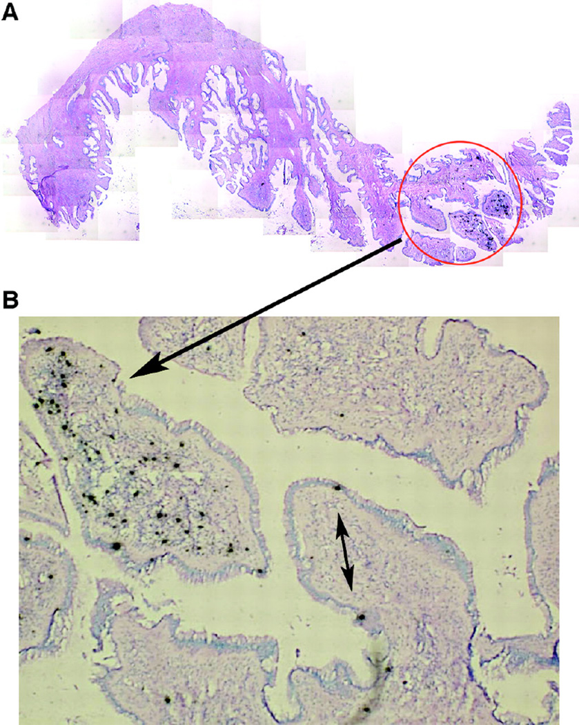
Figure 2. Highly focal productive infection in an endocervix 4 days after intravaginal simian immunodeficiency virus (SIV) inoculation.
A. Montage of images (magnification, ×100) of a single section of endocervix, among 20 sections examined from monkey 31385, in which one focus (encircled) of SIV RNA-positive cells was detectable. B. SIV RNA-positive cells at a higher magnification. The double-headed arrow points to two SIV RNA-positive cells that appear to be intraepithelial lymphocytes. The single-headed arrow points to a focal collection of SIV RNA-positive cells in the endocervical mucosa. SIV RNA was detected by in situ hybridization (ISH) with radiolabeled riboprobes. In the developed radioautographs viewed with transmitted light, the SIV RNA-positive cells appear black because of the large number of silver grains overlying the cell. Original magnification, ×100. Reproduced from: Miller CJ, et. al. Propagation and dissemination of infection after vaginal transmission of simian immunodeficiency virus. J Virol 2005;79(14):9217–9227, with permission.

