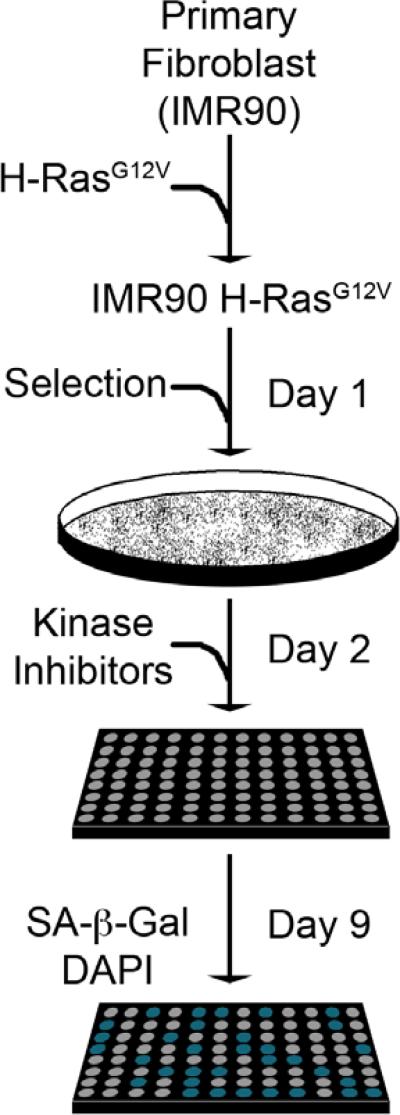
Figure 2. Senescence screen setup and analysis.
Primary fibroblasts (IMR90) were transduced with a H-RasG12V encoding retrovirus over two days (Day -1 and 0). Cells were then selected with puromycin (1 μg/mL) for two days, plated into 96-well plates (1,000 cells/well), and at the end of the second day treated with the kinase inhibitor library at a uniform concentration (250nM). Cells were incubated for seven additional days to allow for senescence and then fixed in a formaldehyde/glutaraldehyde solution. To visualize the SA-β-gal activity and determine cell number, fixed cells were incubated with X-gal and labeled with DAPI.

