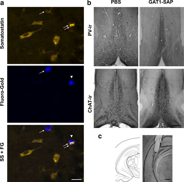
Figure 1.
a, Photomicrographs of hippocampal cells containing SS-ir (top), hippocamposeptal neurons containing FG (middle), and the two images overlaid (bottom). The single arrow shows a SS-ir hippocamposeptal neuron (FG- and SS-ir). The arrowhead points to a hippocamposeptal neuron that is not SS-ir and the double arrows show a SS-ir hippocampal neuron without FG; these two cells are located close together but are not double labeled. Other SS-ir hippocampal neurons that do not contain FG are observed in the top panel. Scale bar, 20 μm. b, Photomicrographs of the MSDB in both PBS- and GAT1-SAP-treated animals. Immunoreactivity for PV and ChAT was used to visualize GABAergic and cholinergic MSDB neurons, respectively. Scale bar (right bottom), 1 mm. c, A brain atlas representation and a photomicrograph of a cresyl violetstained section showing probe placement for the hippocampal microdialysis cannula. Scale bar, 1 mm.

