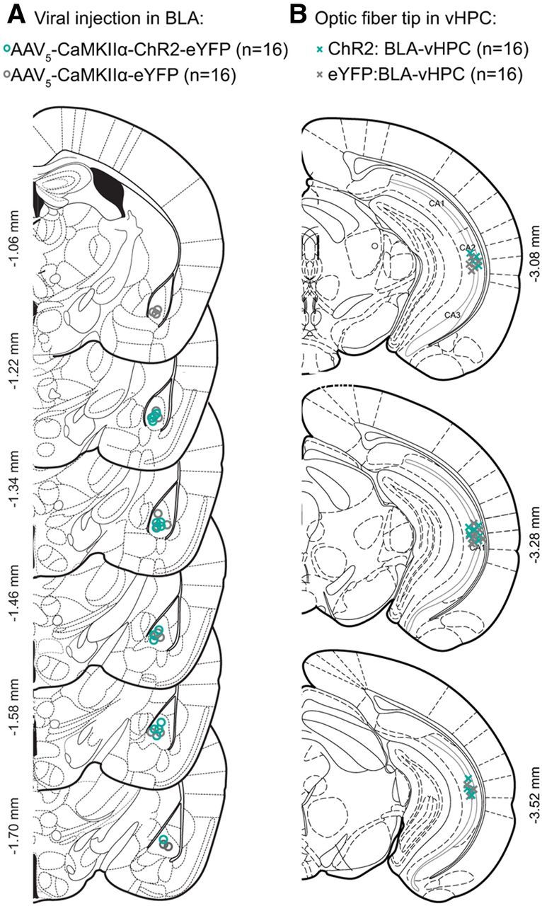
Figure 6.

Histologically verified placements of viral injections and optical fiber tips in ChR2:BLA-vHPC and eYFP:BLA-vHPC groups. A, Coronal sections from bregma of the BLA. Center of the viral injections in the BLA for all the mice injected with ChR2 (n = 16; green circles) and eYFP (n = 16; gray circles). B, Coronal sections from bregma of the vHPC. Location of the optic fibers tip above the pyramidal layer of vHPC for ChR2:BLA-vHPC (green crosses) and eYFP (gray crosses).
