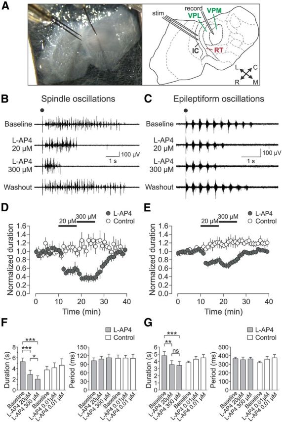
Figure 1.

Group III mGluR activation suppresses evoked thalamic oscillations. A, Horizontal thalamic slice recording setup and schematic. IC, Internal capsule; L, lateral; M, medial; R, rostral; C, caudal. B, C, Representative oscillations, spindle (B) and epileptiform (C), during sequential activation of mGluR4/8 (by 20 μm l-AP4) and mGluR4/7/8 (by 300 μm l-AP4). Electrical stimulus is indicated by black dot. D, Spindle oscillation duration normalized to baseline for l-AP4 treated experimental (n = 5) and control (n = 4) slices. E, Epileptiform oscillation duration normalized to baseline for l-AP4-treated experimental (n = 6) and control (n = 4) slices. F, G, Summary of l-AP4 effects on oscillation duration and period. Spindle oscillations (F) showed effect of epoch on oscillation duration for the experimental group (gray bars; n = 5, F(2,8) = 75.98, p < 0.0001, one-way rANOVA), but not for the control group (white bars; n = 4, F(2,6) = 2.03, p = 0.212). Spindle oscillation duration was significantly decreased further by 300 μm l-AP4 compared with 20 μm l-AP4 (*p < 0.05, Tukey's post hoc test). Epileptiform oscillations (G) also showed an effect of epoch on oscillation duration for the experimental group (gray bars; n = 6, F(2,10) = 17.73, p = 0.0005, one-way rANOVA), but no effect for the control group (white bars; n = 4, F(2,6) = 2.69, p = 0.15). Period did not change for either spindle (experimental group: F(2,8) = 0.42, p = 0.67; control group: F(2,6) = 0.04, p = 0.97, one-way rANOVA) or epileptiform oscillations (experimental group: F(2,10) = 0.21, p = 0.81; control group: F(2,6) = 0.42, p = 0.67). Data are from last 5 min of each epoch. *p < 0.05; **p < 0.01; ***p < 0.001; NS, not significant, Tukey's post hoc test.
