Figure 4.

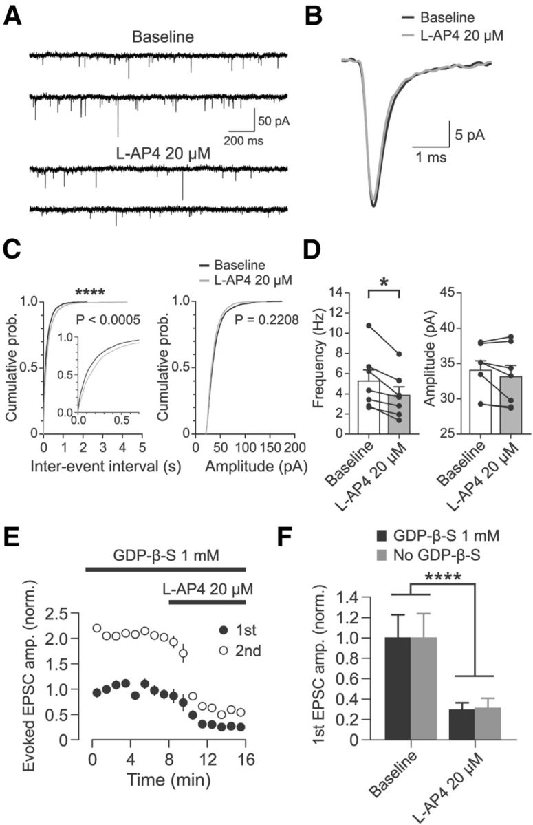
Effect of l-AP4 at cortico-RT synapses is mediated by a presynaptic mechanism. A, Representative partial recording of mEPSCs in a patched RT cell in the presence of 0.5 μm TTX before and after application of 20 μm l-AP4. B, Average mEPSC for experiment shown in A. For each epoch, 200 randomly chosen mEPSCs were peak aligned and averaged. C, Cumulative probability distributions of interevent intervals and amplitudes of mEPSCs before and after application of 20 μm l-AP4 for 200 randomly chosen mEPSCs from each experiment (n = 7, 1400 total events). Interevent intervals were significantly increased (****p < 0.0005, Kolmogorov–Smirnov test), whereas amplitudes were unchanged (p = 0.2208, Kolmogorov–Smirnov test). D, Summary data of l-AP4 resulting in decreased mEPSC frequency (n = 7, *p = 0.02, Wilcoxon signed-rank test) but no change in mEPSC amplitude (n = 7, p = 0.16, Wilcoxon signed-rank test). Individual sample points are median values of all mEPSCs from each experiment. E, Effect of 20 μm l-AP4 on optically evoked cortico-RT EPSC amplitude when postsynaptic G-protein signaling was inhibited by 1 mm GDP-β-S (n = 6). Amplitudes are normalized to mean baseline amplitude of first EPSC. F, Comparison of l-AP4 effect on cortico-RT EPSCs recorded with postsynaptic GDP-β-S (data as in E, n = 6) versus recorded without GDP-β-S (data as in Fig. 3C, n = 8). Although there was clearly a significant main effect of drug epoch (F(1,12) = 33.87, ****p < 0.0001, two-way rANOVA), there was no significant main effect of condition (F(1,12) = 0.022, p = 0.884) or a significant interaction of condition and epoch (F(1,12) = 0.07, p = 0.793). Data are from the last 2 min of baseline and min 3–5 of l-AP4 wash-in.
