
Figure 1.
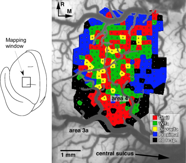
Representation of distal forelimb movements in primary motor cortex (area 4) of a squirrel monkey. Under ketamine sedation, movements were evoked by intracortical microstimulation at each of 321 sites (small white dots) located approximately 250 μm apart. The distal forelimb representation is comprised of digit (red), wrist (w/fa; green), forearm (green) movements, as well as combinations of single-joint movements (yellow). This fractionated pattern of movement representations is due to the intermingling of corticospinal neurons that project to different subsets of motor neurons (Milliken et al., 2013).