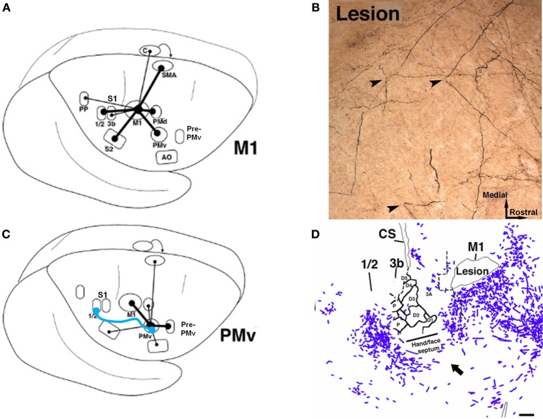
Figure 6.
Rewiring of corticocortical connections after ischemic infarct. (A) In normal healthy squirrel monkeys, the primary motor cortex (M1) has dense reciprocal connections with both the premotor cortex (PMv, PMd, SMA) as well as the primary somatosensory cortex (S1) and the second somatosensory area (S2). (B) In addition to M1, the ventral premotor cortex (PMv) has dense connections with a rostral area called pre-PMv. PMv has moderate connections with S2, but negligible connections with S1. (C) Several weeks after an ischemic infarct in M1, axons originating in PMv can be seen making sharp bends and avoiding the infarct area, as shown in this tract-tracing study. (D) A low-magnification plot of axons within the section show that the axons originating from PMv course around the central sulcus. Substantial terminal bouton labeling (not shown) appears in S1 (areas 1 and 2). The blue line in (B) signifies the de novo pathway that forms after the lesion (Dancause et al., 2005).

