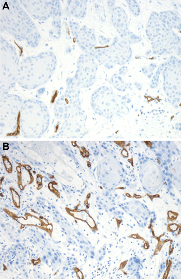
Figure 1.

Representative images of CD34 staining of primary vulvar carcinoma vascularization. (A) Low vascularity (low Chalkley count) and (B) High vascularity (high Chalkley count). Images were taken by a Leica DFC 320 digital camera with a Plan-neofluar 10× objective lens in Axiophot microscope (Zeiss Germany).
