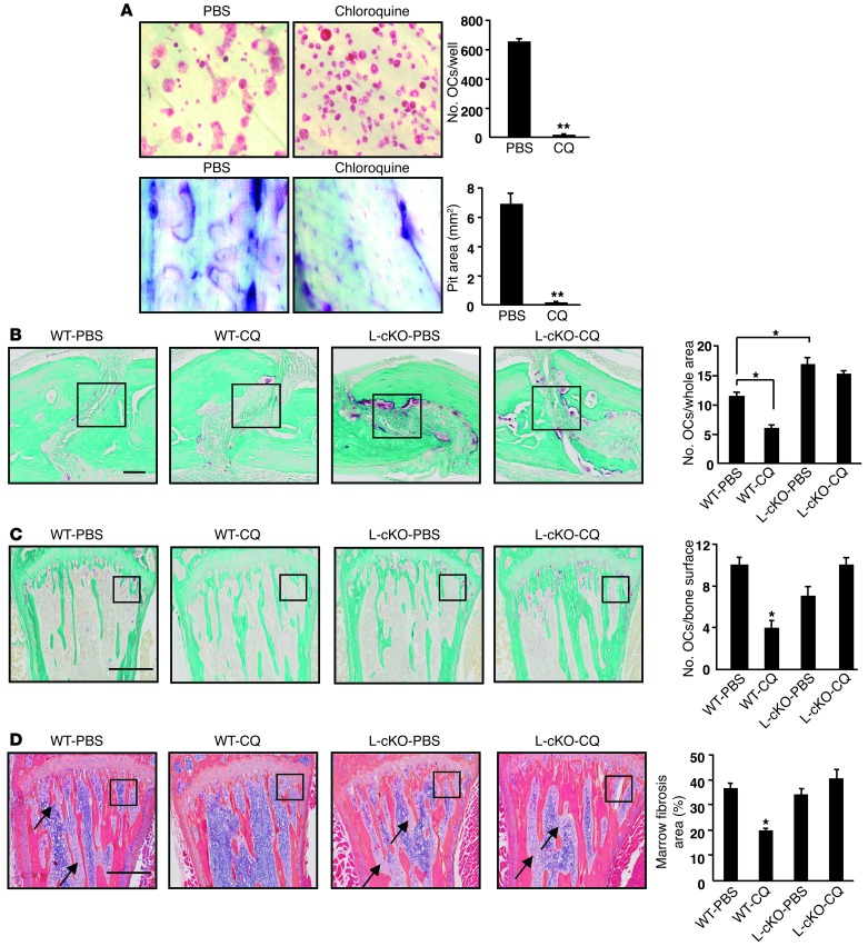
Figure 4. CQ affects OC function in vitro and in vivo.
(A) WT OCPs cultured with RANKL with or without CQ (5 μM) for 9 days. TRAP staining (left panels) and toluidine blue (right panels, to highlight resorption pits). Values are means + SEM of 4 wells. **P < 0.01 vs PBS. Original magnification, ×20. (B–D) Representative TRAP-stained sections and OC numbers in calvarial (B) and tibial (C) sections from 10- to 12-week-old male WT or L-cKO mice treated with CQ (50 mg/kg/d i.p. for 10 days) and given supracalvarial injections of hPTH(1-34 aa) (10 μg/mouse) 4×/d for 3 days beginning on day 7. (D) Representative H&E-stained tibial sections from the mice in C illustrating marrow fibrosis (arrows) and values for percentage of marrow space occupied by marrow fibrosis. Values are means + SEM of 4 mice/group. *P < 0.05. Boxed areas in B–D are illustrated at higher magnification in Supplemental Figure 5. Scale bars: 50 μm (B); 500 μm (C and D).

