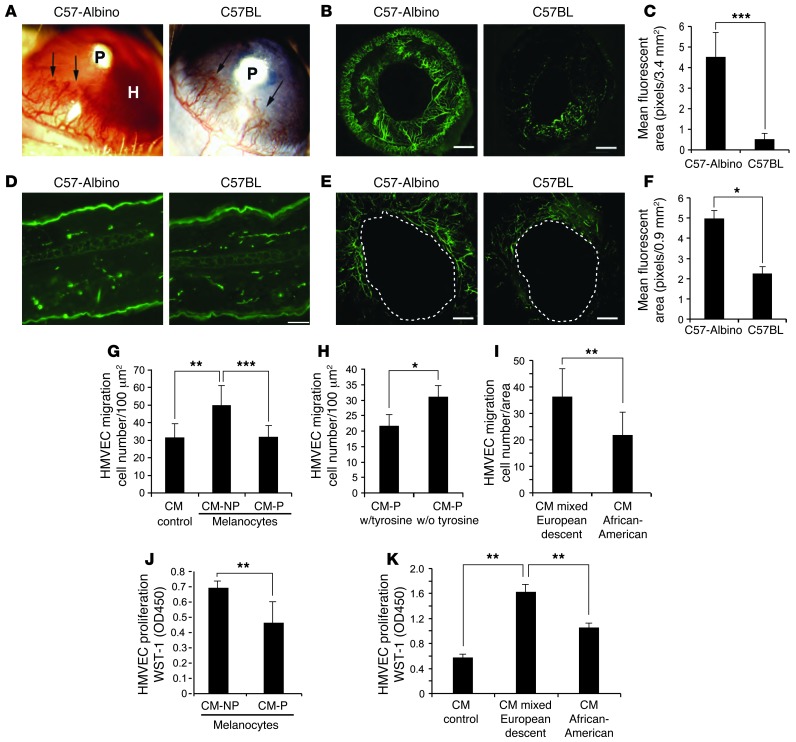
Figure 1. Effect of pigmented and nonpigmented melanocytes on angiogenesis.
(A) Corneal neovascular response in different inbred mouse strains induced by corneal implantation of FGF2 pellets (P) in C57-albino mice (left) with hyphema formation (H) and C57BL mice (right). The experiment included 10 eyes per group. (B) Iris neovascularization visualized with FITC–BS-1 lectin. Scale bars: 500 μm. The experiment included 10 eyes per group. (C) Quantification of iris vessel density of C57-albino versus C57BL. Experiment included 10 eyes per group. (D) Normal skin vessel density in C57-albino and C57BL mice labeled with CD31 antibody. Scale bar: 20 μm. (E) Angiogenesis during wound healing in ear skin. Blood vessels were perfused with FITC-conjugated BS-1 lectin (green) and photographed. Scale bars: 10 μm. The experiment included 5 wounds per group. (F) Analysis of microvessel density in albino ear skin. (G) Migration of HMVECs to CM from albino (CM-NP) and pigmented (CM-P) melanocytes. (H) Migration of HMVECs to CM from pigmented melanocytes grown in the presence and absence of tyrosine. (I) Migration of HMVECs to CM from human melanocytes (mixed European descent and African-American) through Transwells. (J) HMVEC proliferation in response to CM from nonpigmented or pigmented melanocytes for 24 hours; analysis by WST-1 at 450 O.D. (K) HMVECs incubated with CM from human melanocytes (mixed European descent and African-American) for 24 hours, followed by proliferation analysis with WST-1 at 450 O.D. All experiments were repeated at least 3 times. *P < 0.01; **P < 0.001; ***P < 0.0001.

