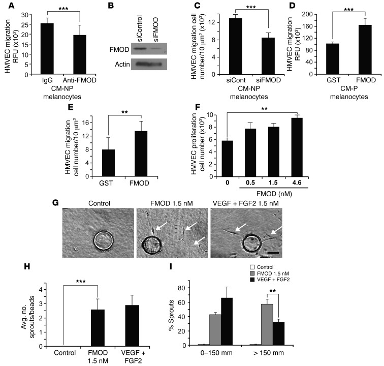
Figure 3. FMOD-induced migration, proliferation and sprouting of HMVECs.
(A) HMVEC migration in response to FMOD-neutralized CM from nonpigmented melanocytes, IgG was used as a control. (B) siRNA-mediated knockdown of FMOD was confirmed by Western blot. β-Actin was used as a loading control. (C) HMVEC migration in response to CM from nonpigmented melanocytes transfected with FMOD-specific siRNA. (D) HMVEC migration was assessed in CM from pigmented melanocytes containing 2 μM recombinant FMOD or GST. (E) HMVEC migration with recombinant FMOD or control-GST. (F) Proliferation of HMVECs with designated concentrations of FMOD was assessed by cell counting. (G) HMVECs are coated onto beads and embedded into a fibrin gel in the presence of medium alone, medium plus 1.5 nM FMOD, or medium plus VEGF-A/FGF2 (1.5 nM each). Arrows show sprouts. Scale bar: 100 μm. (H) Average number of sprouts per bead was not significantly different between 1.5 nM FMOD and positive VEGF-A/FGF2 control. Significant differences were observed between recombinant FMOD and control. (I) Percentage of sprouts with a length less than 150 μm in the presence of 1.5 nM FMOD and 1.5 nM VEGF-A+FGF2 versus those with length greater than 150 μm in the presence of FMOD or 1.5 nM VEGF-A/FGF2. 1.5 nM FMOD increased cell migration over VEGF-A/FGF2. **P < 0.001; ***P < 0.0001.

