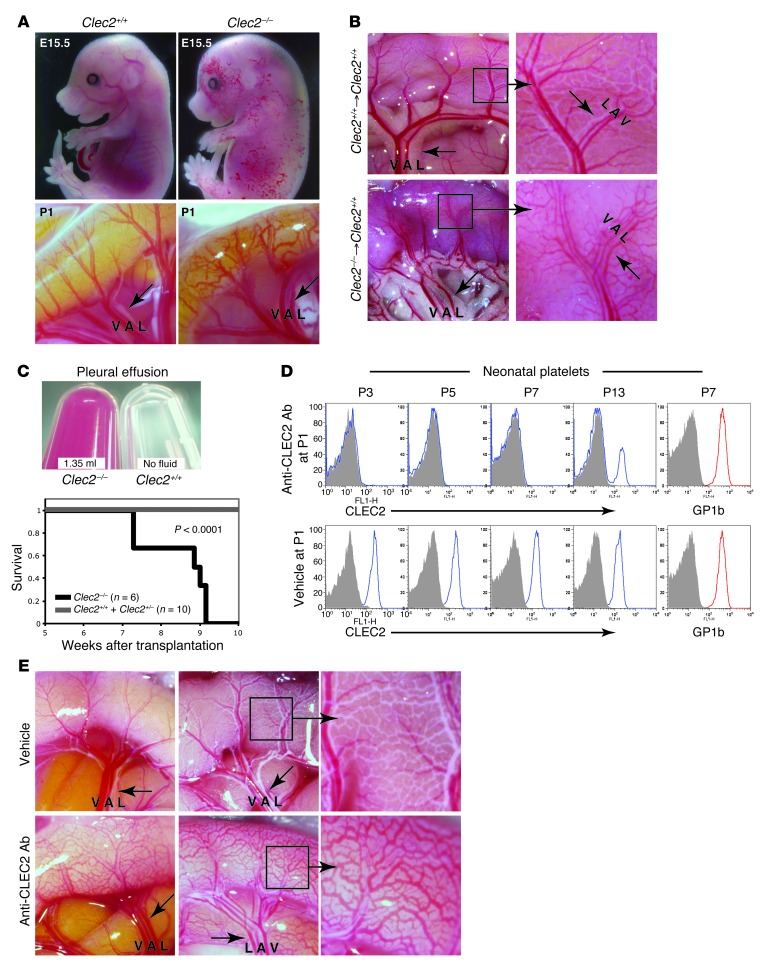
Figure 1. Loss of CLEC2 results in blood-filled lymphatics in the intestine of both perinatal and mature mice.
(A) Genetic deletion of Clec2 resulted in blood-filled lymphatics in the skin at midgestation (top) and in the small intestine at birth (bottom). (B and C) Mature wild-type mice reconstituted with Clec2–/– hematopoietic cells exhibited blood-filled intestinal lymphatics (B) and large, bloody pleural effusions (C). (B) Images of the intestine were obtained 5.5 weeks after transplantation of the indicated hematopoietic cells. Respiratory distress and death were observed in animals with Clec2–/–, but not Clec2+/+ or Clec2+/–, hematopoietic cells. (C) Pleural effusion fluid was obtained at the time of sacrifice. The survival curve of a transplantation cohort is also shown. A large pleural effusion was observed in all 6 Clec2–/– recipients. (D) i.p. injection of the anti-CLEC2 antibody INU1 at P1 resulted in a sustained CLEC2 deficiency state. Shown is flow cytometry of circulating platelets stained with anti-CLEC2 or anti-GP1b antibodies. (E) INU1-mediated CLEC2 deficiency conferred blood-filled intestinal lymphatics. Images of intestine at P5, after a single INU1 injection at P1 (left), or at P14, after injection at P1, P5, and P9 (middle and right). Arrows indicate lymphatic vessels. Boxed regions are shown at higher magnification (enlarged ×3). V, vein; A, artery, L, lymphatic.

