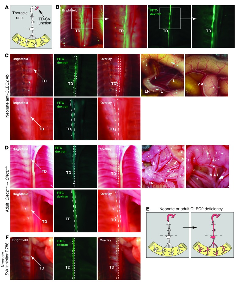
Figure 3. Blood is first observed in the TD after loss of Clec2 in both neonates and mature animals.
(A) Terminal lymphatic network between the intestine and LV junction. Dotted lines denote level of the diaphragm. (B) The TD at P6 was detected adjacent to the vertebral column in the chest by the presence of FITC-dextran after injection into the hindlimb. Boxed regions are shown enlarged at right. (C) Blood was detected in the TD prior to the lymphatics of the mesentery and intestine after loss of CLEC2. Shown are TD (top left) and abdomen (top right) of a P6 wild-type animal injected with anti-CLEC2 antibodies on P1 after FITC-dextran injection. Arrows indicate blood in the TD. Note the absence of blood in lymphatics of the intestine (top) and its presence in the terminal region of the TD near the LV junction (bottom). (D) 12-week-old wild-type mice were lethally irradiated and reconstituted with Clec2–/– hematopoietic cells, and the TD was imaged 3 weeks later. Arrows indicate blood in the TD. Note the absence of blood in lymphatics of the intestine (top right). Blood was first observed in the terminal region of the TD near the LV junction after reconstitution (bottom). (E) Path of blood from the TD to the lymphatics of the mesentery and intestine after induced CLEC2 deficiency. (F) TD after 8 days of fostamatinib treatment of wild-type mice. Blood was not detected in the TD (n = 6).

