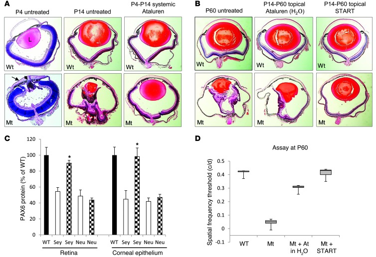
Figure 1. Postnatal treatment of Pax6 mice with ataluren.
(A) Effect of systemic ataluren treatment on mice with the Pax6Sey+/– phenotype. The black arrowhead indicates the lenticular stalk; the black arrow indicates the cornea; and the asterisk indicates the ciliary margin. WT, Pax6+/+; Mt, Pax6Sey/+; L, lens; r, retina. Original magnification, ×5. (B) Histological comparison of 1% ataluren suspension and the START formulation instilled topically in Pax6Sey+/– eyes. Original magnification, ×5. (C) PAX6 protein measurements in the retinas and corneal epithelia from Pax6+/+ (WT), Pax6Sey/+ (Sey), and Pax6Sey–1Neu (Neu) mice. Black bars depict wild-type mice; white bars depict untreated mice; checkered bars depict mice after START therapy. *P < 0.001, n = 6. (D) Box-and-whisker plots comparing maximum spatial frequency threshold of topical ataluren (At.) in H20 and the START formulation. Box-and-whisker plots were prepared showing the 5% and 95% quantiles (whiskers), 25% and 75% quartiles (box), and the median marked by a horizontal line.

