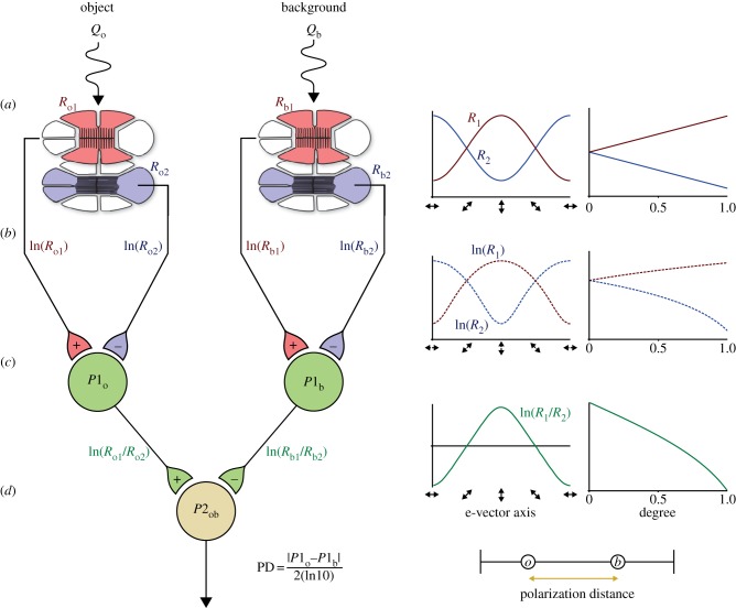
Figure 1.
Schematic of the two-channel polarization model. (a) Polarized light from an object and the background are detected by two receptors in different parts of the eye. Each receptor is composed of two sets of orthogonally oriented receptor cells, vertical (red) and horizontal (blue). Receptor activity is modulated by changes in e-vector axis and degree of polarized light (graphs right). (b) Signals from each set of receptor cells are passed via an opponent system to interneurons, which show inhibitory/excitatory responses. (c) Polarization distance is calculated as a normalized measure of relative P1 interneuron activity (d).

