
Figure 6.
Emerging PSC-Derived Chondrocytes Can Be Enriched Using a Similar Strategy Defined by Chondrocyte Ontogeny in Vivo
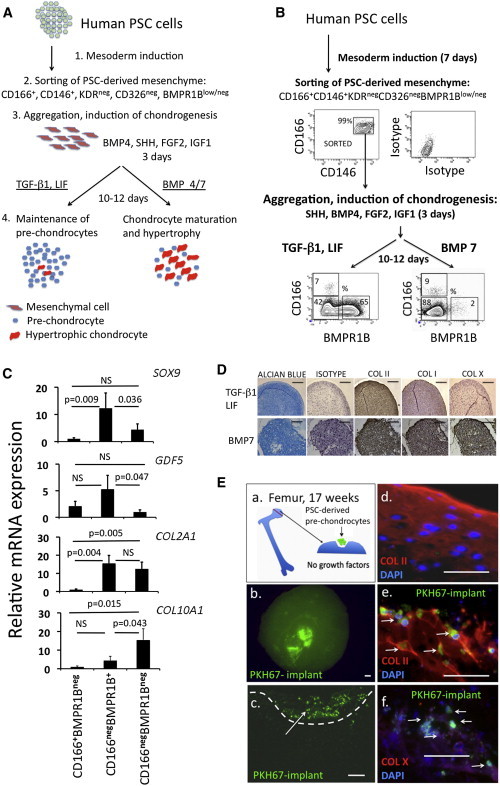
(A) Experimental scheme depicting the generation of PSC-derived chondrocytes following the induction of mesenchyme differentiation.
(B) Immunophenotypic profiles used to isolate prechondrocyte-like and mature chondrocytes generated from PSC-derived mesenchyme after chondrogenic aggregation.
(C) Expression of chondrogenic genes in CD166low/negBMPR1Bneg, CD166low/negBMPR1B+, and CD166+BMPR1Bneg cells isolated from chondrogenic aggregates cultured in the presence of LIF and TGF-β1. CD166low/negBMPR1B+ cells expressed higher levels of prechondrocyte genes compared to the other two populations and showed significantly lower expression levels of COL10A1 than CD166low/negBMPR1Bneg cells (mean ± SD; three independent experiments); NS, not statistically significant.
(D) Chondrogenesis-committed mesenchymal cells cultured in the presence of TGF-β1 and LIF (top row) generate cartilage matrix positive for Alcian blue and collagen II, with minimal levels of collagen I and X, while mesenchymal cell aggregates cultured in the presence of BMP7 show hypertrophic morphology and deposit high levels of collagen X (bottom row). Scale bar shown for images is 100 μm.
(E) a. PSC-derived CD166low/negBMPR1B+ cells were isolated by FACS, labeled with PKH67 green, and deposited into explants from fetal hip joints. b. Explant viewed from above at the time of harvest showing persistence of PKH67-labeled cells after 14 days. c. Transverse section through the explant. d. Uniform Collagen II staining of chondrocytes in the uninjured surface of the explant. e. PSC-derived prechondrocyte-like cells integrate into the injured region and produce collagen II, but not collagen X (f). Scale bar, 50 μm.
See also Figure S5.