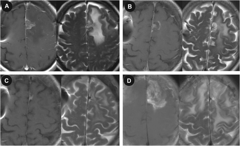
FIGURE 3.

Magnetic resonance imaging (MRI) showing a recurrent glioblastoma treated with focused laser interstitial thermal therapy. A, preoperative T1-weighted MRI with gadolinium and T2-weighted MRI showing a 1.3 × 0.8-cm left frontal lesion. T1-weighted MRI with gadolinium and T2-weighted MRI on postoperative days 1 (B) and 5 (C) and 11 months (D) postoperatively.
