
FIGURE 5.
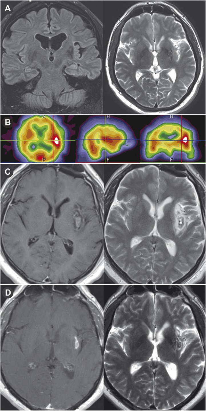
Magnetic resonance imaging (MRI) showing architectural distortion in the left subinsular region that corresponded to an abnormality on the ictal and interictal single-photon emission computed tomography (SPECT) study and was treated with focused laser interstitial thermal therapy. A, preoperative coronal fluid-attenuated inversion recovery MRI and T2-weighted MRI showing a 2.5 × 2.1-cm left subinsular lesion. B, preoperative ictal SPECT study showing increased activity in left insula and temporal lobe. T1-weighted MRI with gadolinium and T2-weighted MRI on postoperative day 1 (C) and 5 months postoperatively (D).