Figure 2.
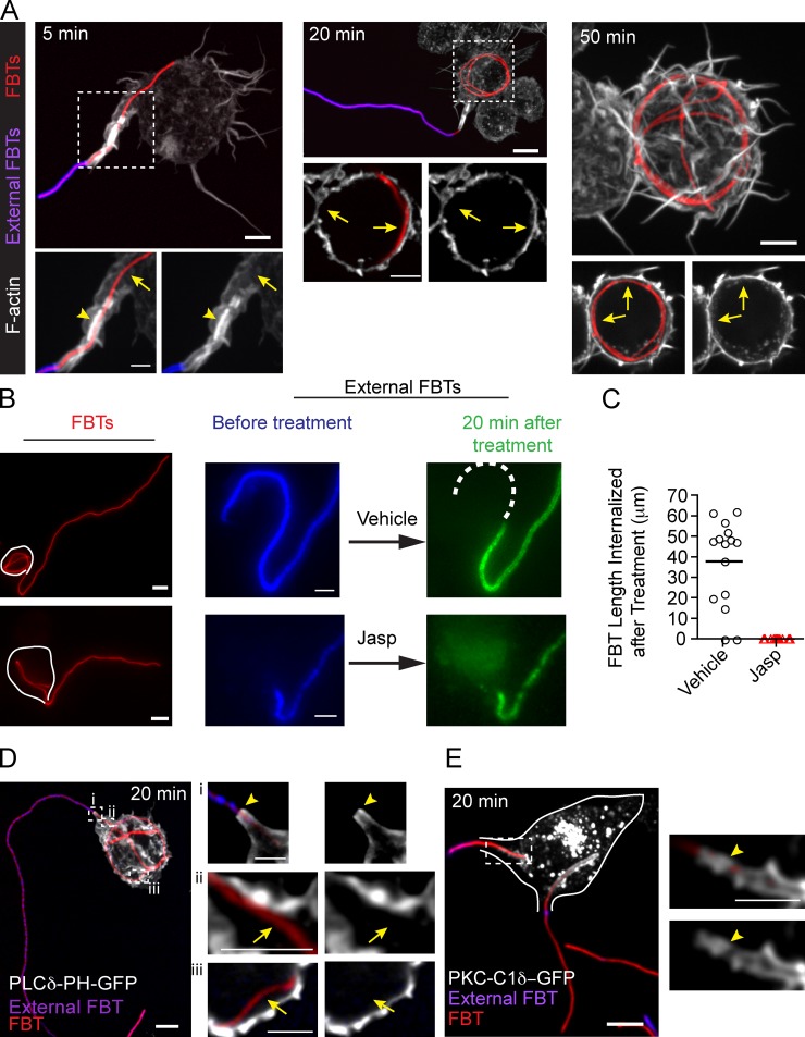
T-PC elongation requires actin turnover. (A) F-actin was enriched in the T-PC (arrowhead) forming the actin jacket but disappeared as the filament continued to get internalized (arrows). Cells were fixed at indicated times after FBT phagocytosis, external FBTs were immunolabeled (pink), and F-actin was stained using Phallodin 488 (white). (B) After 20 min of phagocytosis, external FBTs were immunolabeled (blue) in the cold. Phagocytosis was then allowed to progress in the presence of jasplakinolide (jasp, 20 min). External FBTs were immunolabeled (green) and cells were fixed. Dashed line in the panels to the right indicates the FBT length internalized during the treatment. (C) Lengths of FBTs internalized after the treatment in B. Data shown are FBT lengths from a representative experiment out of three repeats. Lines indicated the means. For the experiment shown, n = 15. (D) PI(4,5)P2 disappeared as FBTs were internalized. Phagocytosis of FBT by cell expressing PLCδ-PH-GFP (white). Cells were fixed after 20 min of phagocytosis and external FBTs were immunolabeled (pink). Right: magnified single planes from framed regions. The presence of PI(4,5)P2 at the T-PCs (arrowheads) and its loss as FBTs are internalized (arrows) are indicated. (E) Diacylglycerol appeared as FBTs were internalized. Phagocytosis of FBT by cell expressing PKC-C1δ–GFP (white). External FBTs are shown in pink. Right: magnified single planes from framed regions. White lines delineate the cell boundary. Bars: (main panels) 5 µm; (magnifications) 2.5 µm.

