Figure 6.
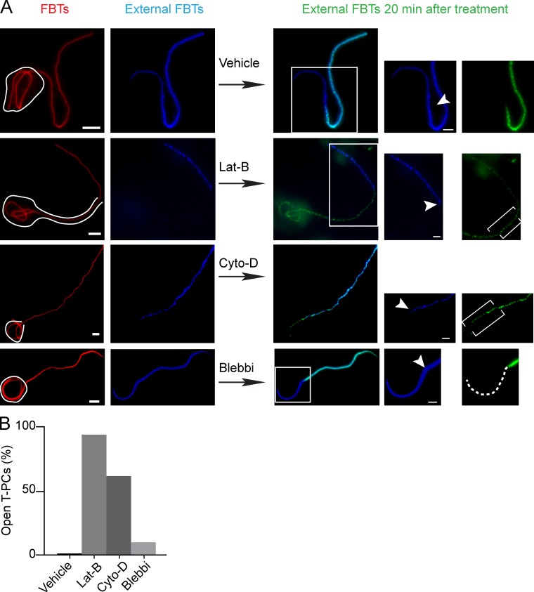
Actin depolymerization disrupts the diffusion barriers at the T-PC. Inhibiting actin dynamics removes the barrier that prevents antibodies from penetrating inside the T-PCs. 20 min after FBT phagocytosis, external segments of FBTs were immunolabeled in the cold (blue). Phagocytosis was allowed to proceed in the presence of latrunculin B (lat-B; 2 µM), cytochalasin D (cyto-D; 10 µM), blebbistatin (blebbi; 100 µM), or vehicle for 20 min and external FBTs were immunolabeled (green). (A) Inhibiting actin remodeling allowed the second round of antibody to penetrate inside the T-PCs (brackets) and label sections of FBTs previously inaccessible (green). Dashed lines in the panels to the right indicate FBT sections that remained inaccessible to the antibodies. (B) Number of T-PCs that lost the diffusion barrier from A. Data shown are percentrages from a representative experiment from three repeats. For the experiment shown, n = 50. Bars: (main panels) 5 µm; (magnifications) 2.5 µm.

