Figure 7.
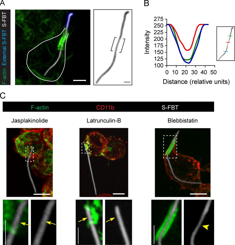
Actin jackets constrict the Salmonella-FBTs. (A) RAW cells were allowed to ingest RFP-Salmonella-FBTs (white) for 10 min. Cells were fixed, external sections of the S-FBTs were immunolabeled (blue), and F-actin was stained (green). Right: higher magnification of the S-FBT (inverted image), showing its constriction by the actin jacket (brackets). Solid line indicates the cell boundary. (B) Constriction of the S-FBT shown in A, as measured by the fluorescence intensity of the indicated regions along the filament. (C) S-FBTs (white) attachment to RAW cells was synchronized in the presence of blebbi (100 µM), jasp (1 µM), or lat-B (2 µM). Cells were fixed 10 min after the initial attachment, and cell membrane labeled with CD11b antibodies (red). F-actin is shown in green. Bottom: higher magnifications of framed regions. Arrowhead indicates S-FBT constriction due to the actin jacket and arrows point to the lack of T-PCs and the constriction of the S-FBTs. All micrographs shown are merged confocal planes (not deconvolved). Bars: (main panels) 5 µm; (magnifications) 2.5 µm.

