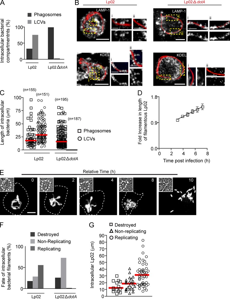
Figure 9.
Filamentous morphology contributes to L. pneumophila survival in macrophages. (A) Number of intracellular RFP-Lp02 or RFP-Lp02-ΔdotA bacteria found in phagosomes and LCVs in macrophages infected for 9 h. Data shown are percentages from a representative experiment from two repeats. For the data shown, n = 150. (B) Representative images of intracellular RFP-Lp02 or RFP-Lp02-ΔdotA bacterial filaments from A. Right: magnified single planes from framed regions. (C) Length of intracellular bacteria and the associated compartment from A. Red lines indicate medians. (D–G) Live-cell video microscopy analysis. Images were acquired using an EM-CCD camera (Hamamatsu Photonics). RAW cells were allowed to internalize RFP-Lp02 or RFP-Lp02-ΔdotA for 5 h, gentamycin was added to the media, and time-lapse video was acquired for infected cells for an additional 15 h to follow the fate of intracellular bacteria. (D) Elongation of intracellular Lp02 filaments followed over time by time-lapse microscopy. Bacterial length was measured at indicated times using the software Volocity. Data shown are lengths from one representative experiment out of 3 repeats where 11 Lp02 filaments were analyzed. (E) Snapshots from time-lapse imaging of RAW cell infected with Lp02 showing the replication of filamentous Lp02 and production of bacillary progeny. Relative time is indicated on the top right corner and infected cell is delineated by dashed line. Insets show DIC images. (F) Infected cells were identified and followed over time to assess the fate of individual bacteria (destroyed, replicating, or nonreplicating). Data shown are percentages from a representative experiment out of two independent experiments. For the data shown, n = 30. (G) Length of intracellular Lp02 measured at the time the time-lapse acquisitions were started and their fate. Red lines indicate medians from three independent experiments. At least 15 intracellular Lp02 were analyzed in each case. Bars: (main panels) 5 µm; (magnifications) 2.5 µm.

