
Figure 3.
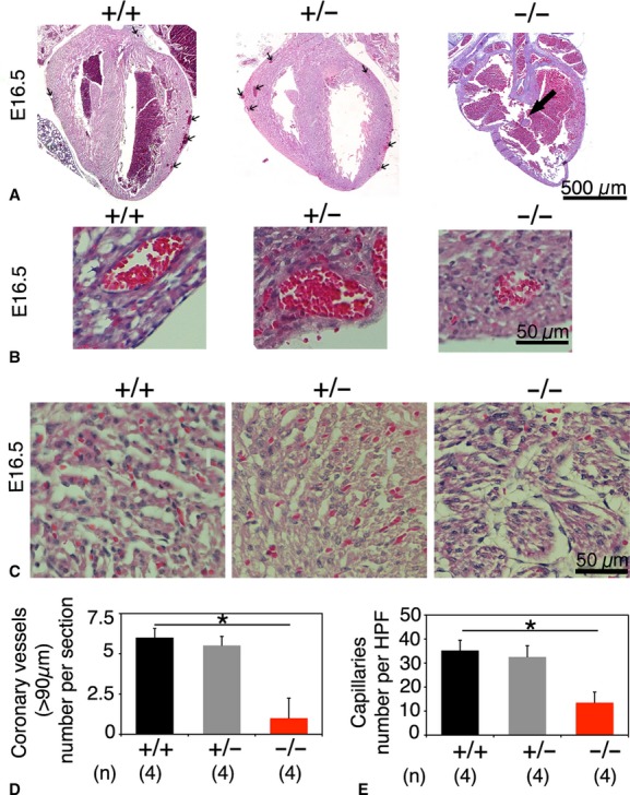
Histological analysis of E16.5 fetal Akt1+/+, Akt1+/−, and Akt1−/− hearts, coronary blood vessels and myocardial capillaries. (A) Some Akt1−/− fetal hearts displayed ventricular septum defects (VSDs; large arrow). Coronary vessels (>90 μm) located beneath the epicardium were rarely present in Akt1−/− fetal hearts (small arrows). (B) Akt1−/− myocardial veins are smaller and have a thinner perivascular wall than Akt1+/+ veins (C) Akt1−/− myocardium exhibited less myocardial capillaries than Akt1+/+ myocardium. (D) Quantification of the amount of coronary vessels (>90 μm) per section. (E) Quantification of the amount of myocardial capillaries per high power field (HPF). (*P < 0.05).