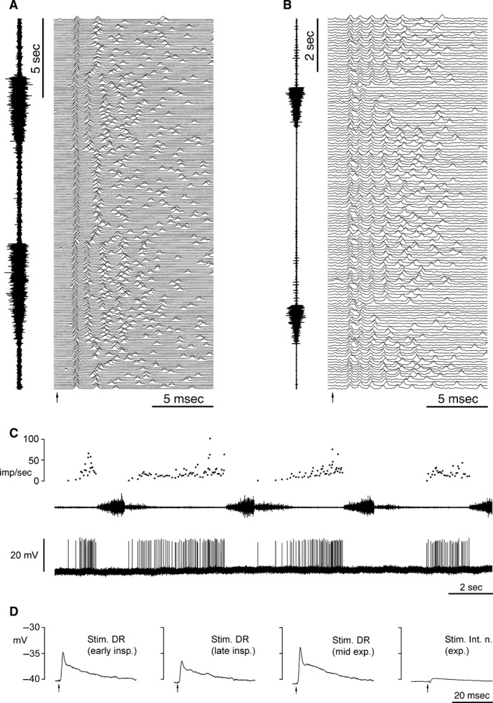
Figure 1.

Electrophysiology of the Renshaw cells. (A, B) Respiratory variation in the responses to nerve stimulation (internal intercostal nerve for both, at the arrows). The intracellular recordings are shown in a raster format in time sequence from bottom to top. (A) Cell B23J, membrane potential −56 to −53 mV, spike amplitude 60–70 mV. (B) Cell B17P, membrane potential −26 mV, spike amplitude 10 mV. For each example, an external intercostal nerve discharge is shown at the right (T5 for A, T6 for B). (C) Spontaneous activity of cell B17P: intracellular recording shown in the bottom trace, firing rate (instantaneous frequency) on the top trace, T6 external nerve discharge in the middle trace. (D) Averaged intracellular responses from cell B18P to nerve stimulation, all within a single respiratory cycle, during early inspiration, late inspiration, and expiration, as indicated. First 3 panels, five sweeps averaged for each panel, stimulation to the dorsal ramus; fourth trace, 41 sweeps, stimulation to the internal intercostal nerve.
