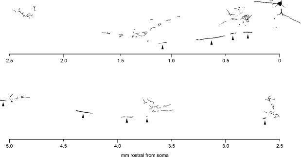
Figure 3.

Drawing of all the labeled fragments in one parasagittal section, from cell B17P. The lower half of the figure should be read as continuous with the top half, aligned by the distance scale. The stem axon, in the white matter, is indicated by arrowheads. Drawing made via a drawing tube attached to the microscope. Dorsal is at the top of the figure, rostral is to the left.
