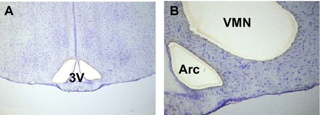
Figure 1.

Laser capture microdissection of the VMN and arcuate nucleus (Arc) from coronal brain sections collected from nonpregnant rats and day 14 pregnant rats. (A, B) Part of a thionin-stained coronal rat brain section used for LMD. (A) Two holes where the Arc has been dissected out on both sides of the third ventricle (3V). The VMN is still intact. (B) Holes where the Arc and VMN have been dissected out on the left side of the third ventricle shown in A. VMN, ventromedial nucleus of the hypothalamus; LMD, laser capture microdissection.
