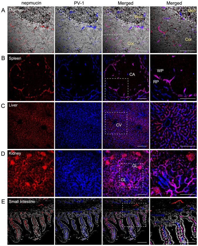
Figure 2. Nepmucin/CD300LG shows heterogeneous expression patterns in distinct compartments of the thymus and spleen.
(A-E) Frozen sections of thymus (A), spleen (B), liver (C), kidney (D), and small intestine (E) were stained with an anti-nepmucin mAb (Alexa Fluor 594; red) and anti-PV-1 mAb (Alexa Fluor 647; blue). Cryosections of the thymus (A) and small intestine (E) were further incubated with Hoechst 33342 (white). In the thymus (A), the cortico-medullary junction is indicated by a dotted line. Med: medulla, Cor: Cortex, CA: central artery, WP: white pulp, RP: red pulp, CV: central vein, GL: glomerulus. Scale Bars, 100 µm.

