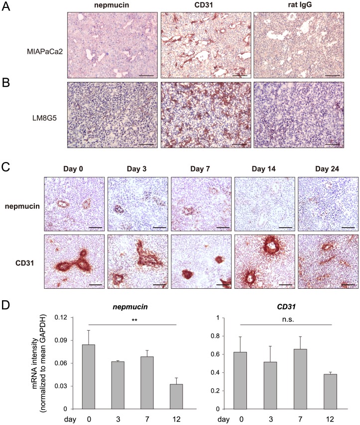
Figure 7. Nepmucin/CD300LG expression is decreased in tumors and tumor-draining lymph nodes.
(A) Human pancreatic adenocarcinoma MIA PaCa-2 cells (1.2×10⁷ cells) were subcutaneously injected into the flank of SCID mice that had been treated with an anti-IL-2Rβ mAb. Eighteen days later, nepmucin expression was examined in the tumor tissues. (B) Nepmucin expression in liver metastatic tumors was examined after LM8G5 osteosarcoma cells (1×10⁶ cells) were intravenously injected into the ileocolic vein of C3H/HeN mice. (C, D) Axillary LNs were harvested from C57BL/6 mice that had been subcutaneously inoculated with 1×10⁵ B16-F10 melanoma cells. The nepmucin and CD31 expressions were examined by immunohistochemistry (C) and quantitative PCR (D) at the indicated time points after inoculation. Data show representative images, n = 3–4 mice per group. **p<0.01, n.s., not significant. Scale bars, 100 µm.

