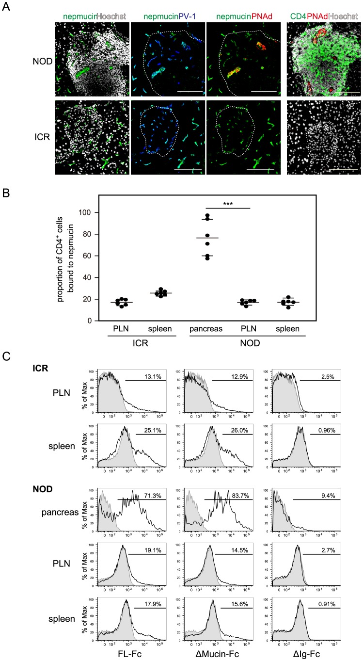
Figure 8. Nepmucin/CD300LG is expressed in NOD pancreas high endothelial venule-like vessels and avidly binds diabetes-associated CD4+ T cells.
(A) The pancreas was collected from 13-week-old female NOD mice and ICR mice. Tissue sections were stained with anti-nepmucin mAb (green), anti-PV-1 mAb (blue), anti-PNAd mAb (red), and Hoechst 33432 (white). Alternatively, a frozen section was stained with anti-CD4 mAb (green), anti-PNAd mAb (red), and Hoechst 33432 (white). The Langerhans’s islets are delineated with dotted lines. Scale bars, 100 µm. (B, C) Lymphocytes prepared from the indicated tissues of NOD or ICR mice (15–19 weeks old) were incubated with admixed nepmucin-human IgG Fc chimeras and PE-conjugated F(ab’)2 anti-human IgG. The proportion of CD4+ T cells bound to full-length nepmucin (FL) is shown in (B), and representative histograms for the binding of nepmucin FL and its mutant (Δmucin; nepmucin mutant lacking the mucin-like domain, and ΔIg; nepmucin mutant lacking the Ig domain) are shown in (C). Data represent the mean ± SD (n = 6 mice per group) from two independent experiments. ***p<0.005.

