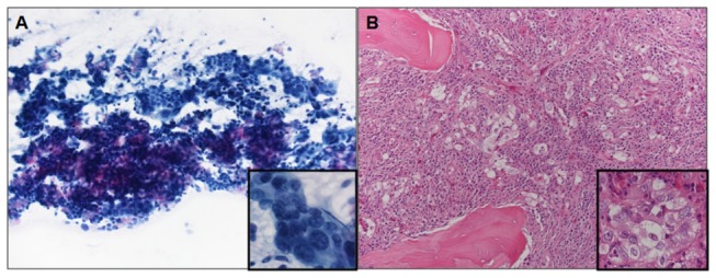
Figure 3. Microscopic evaluation of tumor cellularity in non-small cell lung carcinoma samples.

A) cytology specimen from a 72 year old woman with adenocarcinoma metastatic to a mediastinal lymph node (May Grumwald Giemsa, 200X magnification, inset 600X); the proportion of neoplastic cells in the sample is 35%; DNA analysis was wild type after Sanger sequencing, but NGS showed two EGFR mutations (G721W, R831H) (case 57 of Table 5). B) biopsy specimen from a 65 year old man with adenocarcinoma metastatic to bone (vertebral body) (Hematoxylin and Eosin, 200X magnification, inset 600X); the proportion of neoplastic cells in the sample is 5%; DNA analysis was wild type after Sanger sequencing, but NGS showed the L858R EGFR mutation (case 80 of Table 5).
