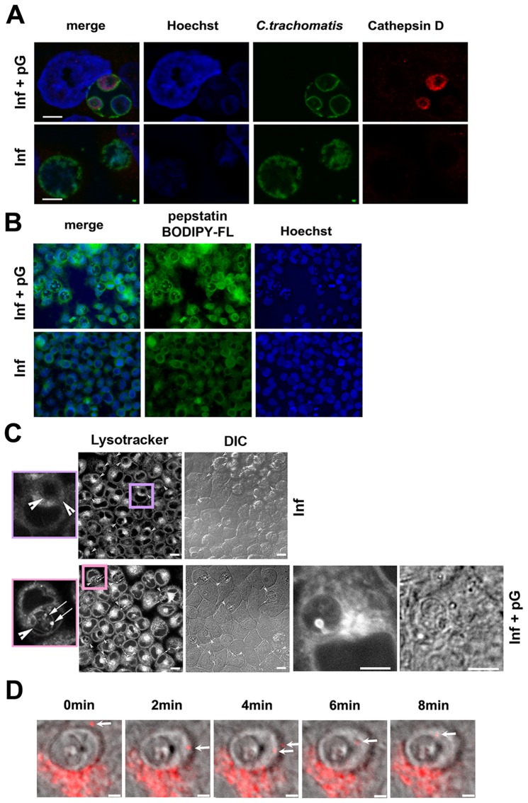
Figure 4. pG induces lysosome entry into bacterial inclusions.
HeLa cells were infected by C. trachomatis serovar L2 and treated with pG at 3 hpi or left untreated. At 24 hpi, cells were either fixed and stained (A and B) or readily observed (C and D). Scale bar: 10 μm. The experiments have been repeated at least 3 times. A- Staining of pG-treated and untreated infected cells as in Figure 1, adding anti-Cathepsin D labelling (red). B- Staining of pG-treated and untreated infected cells using 5 µM BODIPY-pepstatin-FL (green) and Hoechst (blue). C- Cells were incubated with lysotracker for 30 min at 37°C before observation. Lysotracker localizes mainly in inclusions in pG-treated infected cells. Details show strong bright spots in these inclusions, suggesting a lysosome entry. Arrowhead: inclusion, arrow: bright spot. DIC: differential interference contrast. D- Time lapse using a confocal microscope with high resonance scanner. In grey, differential interference contrast, in red, lysotracker. A lysosome (white arrow) enters and stays inside an inclusion in pG-treated infected cells.

