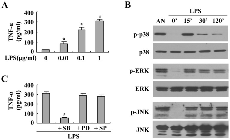
Figure 1. Effect of p38 MAPK on LPS-induced TNF-α production in BMMs.
(A) Cells were treated with different concentrations of LPS for 24 h. Secretion of TNF-α in culture supernatants was detected using ELISA. The results are expressed as the means ± the SEMs of three independent experiments. (B) The cells were treated with LPS (1 µg/ml) for different periods. Protein extracts were subjected to western blotting to detect the phosphorylated and total forms of three MAPK molecules, p38 MAPK, ERK1/2 and JNK, using anti-MAPK antibody. Stimulation with anisomycin (AN, 10 µg/ml for 30 min) was used as a positive control. Representative blots are shown, and the results were verified by at least three independent experiments. (C) Cells were pretreated with specific inhibitors of p38 MAPK (SB203580, 20 µM), ERK1/2 (PD98059, 20 µM) or JNK (SP600125, 20 µM) for 30 min and then exposed to LPS (1 µg/ml) for 24 h. Secretion of TNF-α in culture supernatants was detected using ELISA. The results are expressed as the means ± SEMs of three independent experiments. *P<0.05 compared to the control.

