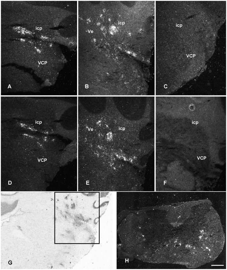
Figure 7. IL-1β and IL-1ra mRNA in brain stem and spinal cord during cr-EAE.
Upper pannels IL-1β mRNA. Middle pannels: IL-1ra mRNA. (A, D) First phase of disease; (B, E) remission; (C, F, H) relapse. (G) anatomical localization of CD68 positive cells during the first disease phase in the lateral brain stem areas including inferior cerebellar peduncles, interpeduncular nuclei, cochlear nuclei, lateral vestibular nucleus and trigeminus. Note the absence of IL-1β and IL-1ra mRNA in the brain stem during the relapse (C, F), while IL-1β and IL-1ra signal is still present in the spinal cord during the relapse (H). Frame in G refers to brain stem areas shown in A–F. Scale bar (A–H) = 500 µm. Icp = inferior cerebellar peduncle; VCP = ventral chochlear nucleus, posterior; Ve = vestibular nucleus.

