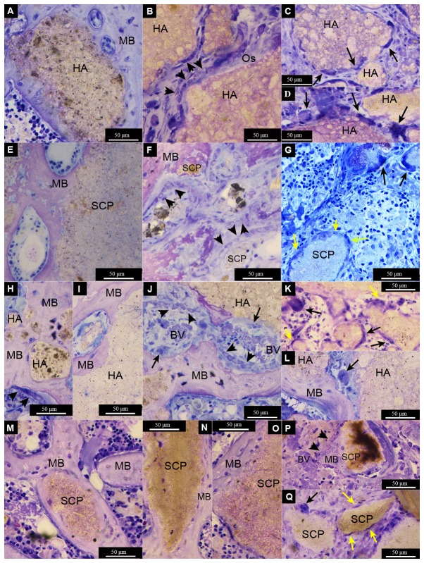
Figure 7. Light micrographs of non-decalcified toluidine blue-stained ground sections of femur defects after 28d of healing.
The defects were created in non-ovariectomised (A-G) or ovariectomised (H-Q) rats. The defects were filled with either hydroxyapatite (HA) (A-D and H-L) or strontium calcium phosphate (SCP) (E-G and M-Q). Mature bone (MB) is formed around and in contact with the granule surface in the four different groups (A = non-OVX HA, E = non-OVX SCP, H and I = OVX HA, M = OVX SCP). Bone formation is clearly visible, with osteoblast seams (indicated by black arrowheads) forming osteoid (Os) appearing directly on some HA granules (B), while mature bone (MB) lined with osteoblasts is observed on the SCP granules (P). A series of osteoblast seams is clearly observed enclosing some granule remnants in the SCP non-OVX defect (F). In the HA OVX defect, signs of bone remodelling are obvious, with the formation of multinucleated cells, blood vessels (BV) and osteoblast seams between the mature bone and granule surface (J). Multinucleated giant cells (indicated by black arrows) were commonly detected at the surface of HA granules (C, D, K and L) and, to a lesser extent, on the SCP granules (G and Q). Spindle-shaped mononuclear cells (macrophage-like cells) (some are indicated by yellow arrows) were also detected in all groups (G, K and Q).

