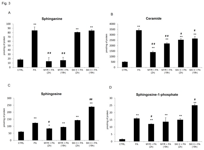
Figure 3. Effects of pharmacological (short and prolonged) inhibition of SPT and SphK1 on intracellular sphingolipids content in L6 myotubes.
L6 myotubes were pre-incubated with 10 μmol/L of myriocin (MYR) or SKI II for 2 h [MYR/SKI II + PA (2 h)] and then incubated with PA (0.4 mM) for next 16 h. In some experiments, L6 myotubes were subjected to incubation with MYR or SKI II for 2 h and during the last 16 h incubation with ceramide inhibitors, cells were also incubated in the presence of palmitate as indicated [MYR/SKI II + PA (18 h)]. (A) Sphinganine (SFA), (B) Ceramide (CER), (C) Sphingosine (SFO), (D) Sphingosine-1-phosphate (S1P). The samples were harvested in ice-cold PBS buffer, and subsequent sphingolipid extraction was performed as described in Methods. Measurement of each treatment was taken as the average of six wells in the same experiments. Data are based on 3 independent determinations. Data are shown as mean ± SEM. *p < 0.05, **p < 0.01 significant difference: control (CTRL) vs. treatment. #p<0.05, ##p < 0.01 significant difference: PA vs. treatment.

