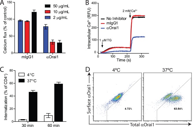
Figure 3. Anti-Orai1 antibody inhibits calcium flux and induces internalization in T cells.
A) Inhibition of thapsigargin-induced calcium flux in Jurkat cells with αOrai1. Data is calculated as percentage of ΔRFU following calcium influx in the absence of antibody addition. B) Representative trace of thaspigargin (TG)-induced calcium influx in Jurkat cells treated with 50 µg/mL αOrai1 or mIgG1 control. C) αOrai1 internalization in purified human CD4+ cells was measured following incubation at 37°C by flow cytometry using 2 µg/mL αOrai1-AF647 and cell surface detection with biotinylated anti-Cy5 followed by streptavidin-BV421. Samples at 4°C were analyzed in parallel as negative controls for internalization. Data is calculated as percentage of total CD4+ cells and is the average of three donors. D) Representative flow cytometry plots of surface bound αOrai1 compared to total internalized and surface αOrai1. Panels A and B are representative of three independent experiments and panels C and D are representative of two experiments.

