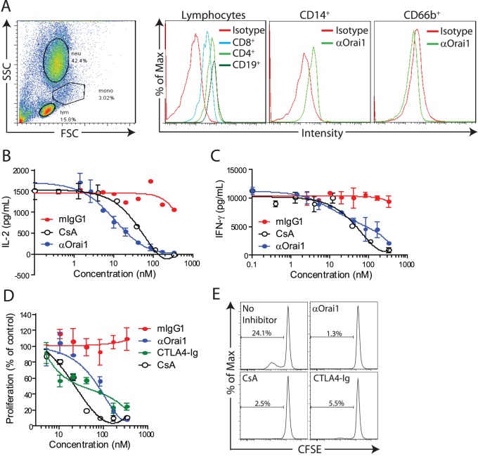
Figure 6. Anti-Orai1 reduces cytokine production by RA synovial fluid cells.
A) Representative forward and side scatter of synovial fluid cells (SFCs) with gating of lymphocytes (lym), monocytes (mono), and neutrophils (neut) and surface staining of Orai1 on CD4+, CD8+, and CD19+ lymphocytes, CD14+ monocytes, and CD66b+ neutrophils from RA synovial fluid. B) IL-2 and C) IFN-γ secretion from αCD3/αCD28 costimulated SFCs following 40 hour-culture in the presence of mIgG1 isotype control, αOrai1, or cyclosporine A (CsA). D) Proliferation of RA patient PBMCs treated with SEB in the presence of control mIgG1, anti-Orai1, CTLA4-Ig, or cyclosporine A (CsA). Data is calculated as percentage of CFSE-diluted cells in the absence of inhibitor. E) Representative CFSE dilution traces of SEB-induced proliferation of RA PBMCs in the presence of 333 nM inhibitor. All panels are representative of two independent experiments.

