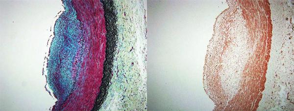
Figure 3.

Representative inflammatory atheroma showing ICAM-1 expression. Movat’s staining on the left (red=smooth muscle cell or endothelial cell, black=elastic fibers, blue-green=extracellular matrix) and custom ICAM-1 staining on the right (brown=ICAM).
