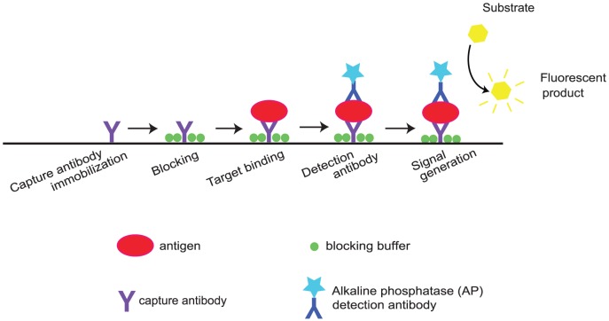
Figure 1. Schematic workflow depicting sequential molecular binding events of the sandwich ELISA.
Within each reaction chamber, the capture antibody is adsorbed on the reactive surface followed by surface passivation by a blocking buffer. Upon target binding to the capture antibody, alkaline phosphates (AP)-tagged detection antibody specific to the antigen is added. Addition of fluorescent substrate (PNPP or p-nitrophenyl phosphate for the traditional well format, and Attophos for the micofluidic format) activated by AP generates detectable fluorescent signal, indicating successful binding events.

