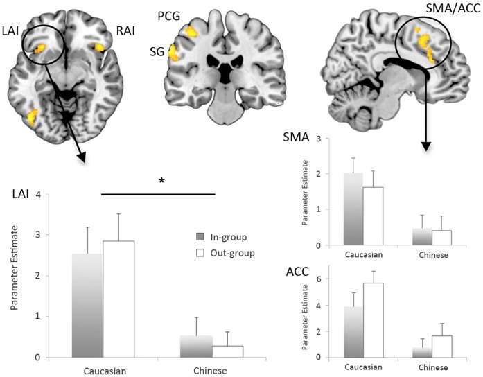
Figure 3. Activation results in the fMRI task.
Significantly greater activitation when observing painful versus non-painful touch was found in the left anterior insula (LAI), right anterior insula (RAI), postcentral gyrus (PCG), supramarginal gyrus (SG), and in the supplementary motor area and anterior cingulate (SMA/ACC). Significant differences when viewing painful touch in Caucasian versus Chinese faces were found only in the left anterior insula, with no differences between In-Group versus Out-Group faces (cluster-level PFWE<0.05; parameter estimates plotted below left). Similar effects in the SMA/ACC failed to reach significance but are shown here for comparison (at voxel-level Puncorrected<0.001; parameter estimates below right).

