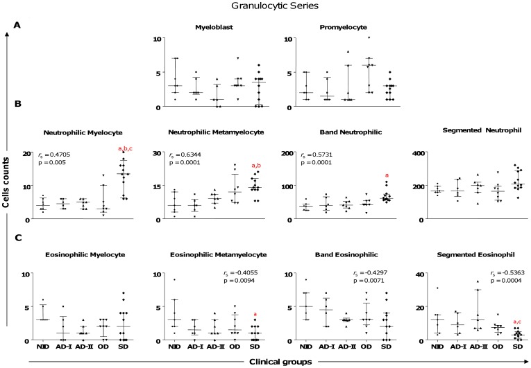
Figure 2. Profiles of the precursors of granulocytic lineage cells in dogs naturally infected by Leishmania infantum categorized according to clinical status.
Animals categorized according to their clinical status into asymptomatic I (AD-I, n = 6), asymptomatic II (AD-II, n = 7), oligosymptomatic (OD, n = 8), symptomatic (SD, n = 12), or noninfected (NID, n = 7) groups. Significant differences at p<0.05 are identified by the letters a, b, and c in comparison to NID, AD-I, and AD-II respectively. The results are expressed in graphics as scattering of individual values and are also shown as median and interquartile range values. Spearman’s rank correlation (r s and p values) are also shown.

